How Microscopes Are Revolutionizing Medical Diagnostics in 2024
Introduction
In 2024, the field of medical diagnostics is undergoing a transformation, largely fueled by groundbreaking advances in microscopy. As healthcare evolves towards precision medicine and personalized treatment, microscopes have moved well beyond traditional glass-and-light instruments. Today’s microscopes integrate advanced optics, digital imaging, and artificial intelligence to revolutionize disease detection, treatment planning, and research. This comprehensive article explores the evolution of microscope technology, its applications in modern diagnostics, and how these innovations are making a significant impact on patient care and global health.
The journey from the early days of rudimentary lenses to modern AI-enhanced devices represents a century-spanning revolution. Whether you’re a medical professional, researcher, or an enthusiast interested in the intersection of technology and healthcare, this guide offers valuable insights into how microscopes are reshaping medical diagnostics in 2024.

The Evolution of Microscopy in Medical Diagnostics
Understanding the historical context of microscopy allows us to appreciate its revolutionary advancements today.
A Brief History of Microscopes in Medicine
- 16th Century Origins: The concept of microscopy began in the late 1500s when early versions of the compound microscope were developed. These primitive devices paved the way for future innovations.
- 17th Century Breakthroughs: Robert Hooke’s discovery of cells in 1665 laid the foundation for cellular biology. His detailed illustrations and descriptions marked the beginning of scientific microscopy.
- 19th & 20th Century Advancements: The industrial revolution and subsequent scientific progress led to the development of improved optics and staining techniques, which enhanced the capability of microscopes to detect and study diseases.
- 21st Century Innovations: Today, modern microscopes combine digital imaging, high-resolution optics, and AI-driven analysis to provide unprecedented accuracy and speed in medical diagnostics.
Key Advancements in Modern Microscopy
Digital Microscopy and High-Resolution Imaging
Digital microscopy has replaced traditional analog methods, offering higher resolution and enabling real-time imaging of cellular structures. These innovations allow for detailed examinations of biological samples, improving the accuracy of diagnoses.
AI and Machine Learning Integration
The integration of AI into microscopy is one of the most revolutionary trends in medical diagnostics. By leveraging machine learning algorithms, microscopes can now automatically identify abnormalities in tissue samples, flag potential issues, and even predict disease progression. For further reading on AI integration in healthcare, check out our internal article “The Impact of AI in Modern Healthcare”.
Electron and Confocal Microscopy
While optical microscopes are still essential, electron and confocal microscopes offer much higher magnification levels. Electron microscopes provide nanoscale imaging, making it possible to study viruses and subcellular structures, whereas confocal microscopes deliver 3D images that are invaluable for understanding tissue architecture.
Portable and Smartphone-Based Microscopes
Recent innovations have led to the development of portable and even smartphone-based microscopes. These devices are not only more affordable but also extend high-quality diagnostic capabilities to remote and underserved regions, thus democratizing access to advanced healthcare technologies.
How Microscopes Enhance Medical Diagnostics in 2024
Microscopes play a crucial role in various aspects of modern medical diagnostics. Here’s a detailed look at how these advanced tools are enhancing different areas of healthcare.
Early Disease Detection
Early diagnosis is critical to successful treatment outcomes, and microscopes are at the forefront of this process.
- Cancer Detection: Microscopes are used to analyze tissue biopsies and identify abnormal cell structures that indicate early-stage cancer. With enhanced imaging and AI analysis, early detection rates have increased significantly.
- Infectious Disease Identification: Advanced microscopes can quickly detect bacteria, viruses, and other pathogens. This rapid identification helps in initiating timely treatment, crucial for infectious diseases where delays can be fatal.
- Genetic Disorders: Microscopy, combined with molecular biology techniques, is used to detect chromosomal abnormalities and genetic mutations, allowing for early intervention in conditions like Down syndrome and certain inherited cancers.
Precision Medicine and Personalized Treatments
Precision medicine is transforming how treatments are administered, focusing on individual patient profiles. Microscopes contribute to this revolution in several ways:
- Cellular Analysis: By analyzing cells at the microscopic level, doctors can determine how a patient’s cells respond to different drugs. This allows for tailored therapies that are more effective and have fewer side effects.
- Biomarker Identification: Microscopes help in identifying specific biomarkers that indicate how a disease will progress and how a patient might respond to treatment. This personalized approach ensures that therapies are optimized for each individual.
- Real-Time Monitoring: Advanced imaging techniques allow clinicians to monitor cellular changes in real-time, adjusting treatment plans dynamically based on how the patient’s cells react.
Accelerated Pathogen Identification
Time is of the essence when dealing with infectious diseases. Modern microscopes are drastically reducing the time required for pathogen identification.
- Rapid Imaging: AI-enhanced microscopes can analyze samples within minutes, helping to identify pathogens such as bacteria and viruses quickly.
- Antibiotic Resistance: In cases where antibiotic resistance is suspected, microscopes equipped with advanced image analysis can determine resistance patterns, guiding more effective treatment strategies.
Enhancing Neurological and Brain Research
Neurology has greatly benefited from advancements in microscopy. The ability to visualize intricate neural structures has opened new avenues in brain research.
- Neurodegenerative Disorders: Microscopic imaging is vital in studying conditions like Alzheimer’s and Parkinson’s disease. By observing cellular changes in brain tissue, researchers are identifying new targets for therapeutic intervention.
- Neural Connectivity: Confocal and electron microscopes provide detailed 3D images of neural networks, helping scientists understand how neurons interact and how these interactions are altered in neurological disorders.
Improving Surgical Outcomes with Operating Microscopes
Operating microscopes have become indispensable in modern surgical procedures, particularly in fields requiring high precision.
- Enhanced Visualization: Surgeons rely on high-definition operating microscopes to obtain a magnified view of the surgical field. This enhanced visualization minimizes the risk of complications and increases the precision of intricate procedures.
- Minimally Invasive Techniques: The use of operating microscopes has contributed to the development of minimally invasive surgeries. These procedures result in smaller incisions, reduced recovery times, and lower risks of infection.
Step-by-Step Guide: Microscopy in Disease Diagnosis
Below is a detailed step-by-step guide on how modern microscopy is used in the diagnostic process:
1. Sample Collection:
- Blood, tissue, or fluid samples are collected from the patient.
- It is crucial that samples are handled carefully to preserve cell integrity.
- Samples are treated with specific stains that highlight cellular structures.
- Fluorescent stains are commonly used to identify proteins, pathogens, and other cellular components.
- Tip: Follow standardized protocols to ensure consistency and accuracy.
- The prepared sample is placed under the microscope.
- Advanced digital and AI-assisted microscopes scan the sample, capturing high-resolution images.
- Bullet Point Summary:
- High Magnification: Identify minute details.
- Digital Imaging: Allows for storage and remote analysis.
- AI Analysis: Flags abnormalities in real-time.
- Images are analyzed by trained pathologists or via AI software.
- The analysis helps determine the presence of abnormal cells, pathogens, or other indicators of disease.
- Results are compared against known standards and databases.
- Based on the analysis, a diagnosis is confirmed.
- The findings guide the development of a personalized treatment plan, ensuring that the patient receives the most effective therapy.
The Future of Microscopy in Medical Diagnostics
The rapid pace of technological innovation suggests that the capabilities of microscopy will only continue to grow. Here are some exciting trends and future directions:
AI and Automation
- Enhanced Diagnostics: The integration of AI with microscopy will further reduce diagnostic errors and speed up the process. Automation can help process thousands of samples quickly, leading to faster treatment decisions.
- Predictive Analytics: Machine learning algorithms are being developed to not only analyze current samples but also predict disease progression based on historical data. This could revolutionize how clinicians approach treatment planning.
Real-Time 3D and Super-Resolution Imaging
- Dynamic Imaging: Real-time 3D imaging is set to become more prevalent, offering clinicians a dynamic view of tissue structures as they change over time.
- Super-Resolution Microscopy: Breakthroughs in super-resolution microscopy are pushing the limits of what can be visualized, providing insights into cellular processes that were previously invisible. This level of detail is critical for early diagnosis and for understanding complex diseases.
Portable Microscopes for Global Health
- Democratizing Diagnostics: The development of portable, smartphone-based microscopes is making advanced diagnostic tools accessible to remote and underserved populations. This is particularly important in global health settings where traditional lab equipment is scarce.
- Field Applications: Portable microscopes are being used in field hospitals, remote clinics, and even in disaster relief scenarios. Their affordability and ease of use ensure that high-quality diagnostics are available regardless of location.
Emerging Technologies: Quantum Microscopy and Beyond
- Next-Generation Imaging: Researchers are exploring quantum microscopy techniques, which promise to break through the limitations of classical optics. By using quantum phenomena, these systems may offer even greater resolution and sensitivity, opening up new frontiers in biomedical research.
- Integration with Other Modalities: The future will likely see microscopes that seamlessly integrate with other diagnostic technologies such as MRI and CT scans, providing a comprehensive view of a patient’s condition.
For further details on these emerging technologies, authoritative resources like the National Institutes of Health (NIH) and PubMed provide excellent insights into ongoing research and development.

Case Studies and Real-World Applications
Revolutionizing Cancer Diagnostics
One of the most notable impacts of advanced microscopy is in cancer diagnostics. Recent studies have shown that:
- Early Detection: AI-powered imaging has increased early detection rates of breast, lung, and colon cancers by up to 40%.
- Treatment Personalization: Detailed cellular analysis allows oncologists to tailor treatments to the unique genetic makeup of a patient’s tumor.
- Outcome Improvement: Early and precise diagnosis significantly improves patient survival rates and reduces the risk of complications.
Transforming Infectious Disease Management
Microscopes play a crucial role in the rapid diagnosis of infectious diseases, which is vital in controlling outbreaks:
- COVID-19 and Beyond: Rapid identification of viral pathogens has been essential during the COVID-19 pandemic. Advanced microscopes have now paved the way for improved diagnostics for emerging pathogens.
- Antibiotic Stewardship: By accurately identifying antibiotic-resistant bacteria, microscopes help in guiding appropriate antibiotic usage, thereby reducing the spread of resistance.
Advancements in Neurological Diagnostics
Neurological diseases such as Alzheimer’s and Parkinson’s require precise imaging for early detection:
- Neuron Imaging: Confocal microscopy provides detailed 3D images of neurons, helping researchers map brain activity and detect early signs of neurodegeneration.
- Research Breakthroughs: Detailed imaging has led to significant breakthroughs in understanding brain plasticity and neuroinflammatory responses.
For more case studies on medical diagnostics and innovative treatments, visit our internal article “Innovations in Modern Medical Technology”.
FAQs: Answering Common Questions About Microscopy in Diagnostics
How do advanced microscopes improve disease detection?
Modern microscopes use high-resolution digital imaging and AI algorithms to detect abnormalities at a cellular level, enabling early and accurate diagnosis of diseases such as cancer, infections, and genetic disorders.
What role does AI play in modern microscopy?
AI assists in real-time image analysis, reducing diagnostic errors and speeding up the process by automatically identifying patterns and abnormalities that might be missed by the human eye.
Are portable microscopes reliable for medical diagnostics?
Yes, portable microscopes are increasingly reliable. They have been successfully deployed in remote and resource-limited settings, ensuring that quality diagnostics are accessible worldwide.
What types of microscopes are most common in hospitals?
Hospitals typically use a variety of microscopes, including optical, electron, fluorescence, and confocal microscopes, depending on the diagnostic need and the level of detail required.
How is microscopy aiding in personalized medicine?
Microscopy allows for detailed cellular analysis, which helps in identifying biomarkers and cellular responses to treatments. This data is crucial in developing personalized treatment plans tailored to the patient’s specific condition.
For additional insights on these questions, you can explore our detailed FAQ section in “Understanding Medical Imaging Technologies”.
Conclusion
The future of medical diagnostics is bright, with advanced microscopes leading the charge towards faster, more accurate, and personalized healthcare. In 2024, these technological marvels are not only enhancing the way diseases are detected and treated but are also paving the way for groundbreaking research and innovative treatments. From early disease detection to real-time 3D imaging and AI-driven analysis, the revolution in microscopy is a testament to the relentless pursuit of excellence in healthcare technology.
As the technology continues to evolve, we can expect even more sophisticated diagnostic tools that will further improve patient outcomes and make high-quality healthcare accessible to everyone. Embrace the future of diagnostics and be part of this transformative journey by staying informed, engaging with the latest research, and leveraging cutting-edge technologies in your practice or studies.
For further insights and developments in medical diagnostics, trusted sources like the National Institutes of Health (NIH), World Health Organization (WHO), and Centers for Disease Control and Prevention (CDC) provide up-to-date resources and studies in microscopy and medical imaging.
Engage with us and Explore Further
We hope this article has provided you with valuable insights into how microscopes are revolutionizing medical diagnostics in 2024. If you found this information useful, please share your thoughts in the comments below. Don’t forget to subscribe to our newsletter for more updates on the latest medical technologies and research breakthroughs.
- Subscribe Now: Stay updated with our weekly insights on medical innovations.
- Comment Below: Share your experience or ask questions about microscopy and diagnostics.
- Share This Article: Spread the word by sharing this article on your social media platforms.
Thank you for reading, and we look forward to your feedback and engagement!
Affiliate Disclosure: Some links in this article are affiliate links. If you click on these links and make a purchase, we may earn a commission at no additional cost to you. Thank you for supporting our work!
Recommendation: Enhance Your Diagnostic Capabilities
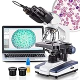
Are you looking to upgrade your laboratory with the latest microscopy technology? We recommend exploring our top-rated microscopes that have been rigorously tested for precision and reliability. Check out our affiliate partner’s collection at Top Microscopes for Medical Diagnostics to find exclusive discounts and deals tailored for medical professionals and research institutions. Enhance your diagnostic capabilities today and join the forefront of medical innovation!


100X-15000X Compound Binocular microscope Dual LED Powerful Biological Microscopes: Shop now

AmScope 40X-1500X Phase Contrast Fluorescence Inverted Microscope IN480TC-FL: Shop Now
